A Behold Interior a Termite’s Gut and More Award-Worthwhile Photos of the Diminutive World
A total world that we never seek for exists under the lens of a microscope. For the past 11 years, talented folks absorb captured dinky existence on movie for the Nikon Little World in Motion Photomicrography Competition. The winning entries introduced a tiny world into fascinating level of curiosity by shooting video photos or digital time-lapse pictures, with the support of a microscope. The entries were then judged by a panel of consultants in the photomicrography and pictures enviornment.
This international competitors has persevered to creatively mix science and technology since 1975. In 2011, Nikon added a video class to its photomicrography competitors, titled Little World in Motion. Since then, the dwell five movies that cleverly showcase the minute world under the lens had been awarded first by fifth function every 365 days.
Listed below are the dwell five winning entries of 2021 that captured a tiny world in motion:
#1. Microfauna in a Termite Tummy
(Courtesy of Nikon Little World/Fabian J. Weston)
Fabian J. Weston, from Pennant Hills, Contemporary South Wales, Australia took home the first-function prize for shooting dinky animals identified as microfauna in a termite’s belly. In his video displayed at 10x, 20x and 40x magnification, you would also seek for that these tiny creatures play a prime function in contributing to termite nutrition. They assist by digesting cellulose, a termite’s essential invent of food.
#2. A Human Micro-Tumor Types and Spreads
(Courtesy of Nikon Little World/Stephanie Hachey and Christopher Hughes)
In this 10-day time lapse that shows an engineered microtumor forming and metastasizing, molecular biologists Stephanie Hachey and Christopher Hughes from the College of California created a specialized atmosphere of their lab that used to be managed for CO2 and humidity. The scientists frail confocal and fluroescence imaging of their work, a technique that allowed them to obviously illuminate a organic course of that is seldom seen. Right here, at 10x magnification, blood vessels (in pink) are supporting the increasing tumor (in blue).
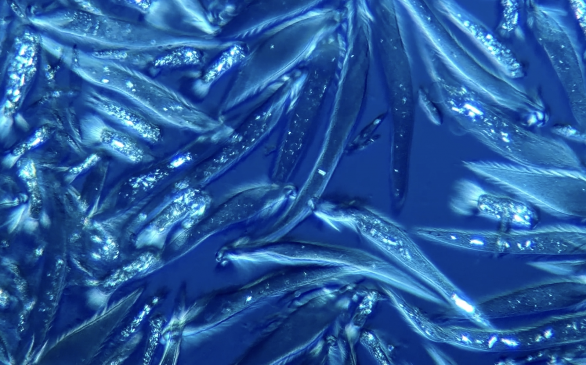
#3. A Water Flea Mama Giving Birth to Miniature Cubs
(Courtesy of Nikon Little World/Andrei Savitsky)
Look a couple of tiny water fleas (Daphnia pulex) swimming away heavenly seconds after their mom gives delivery to them. Andrei Savitsky from Cherkassy, Ukraine captured this unbelievable video, magnified at 4x. A technique called darkfield microscopy lluminated these minuscule aquatic creatures and made them seen of their pure habitat.
#4. Nerve Fibers in Motion After Crossing the Midline of the Central Apprehensive Machine
(Courtesy of Nikon Little World/Alexandre Dumoulin)
Dive deep into the central worried system and glimpse commissural axons (nerve fibers) in motion as they injurious the midline, the worried system’s organizational center. Alexandre Dumoulin from the College of Zurich frail confocal imaging at 40x magnification to highlight these nerve fibers that converse knowledge and join the two aspects of bilateral animals.
#5. An Infected Mosquito Preys on Malaria Parasites
(Courtesy of Nikon Little World/Sachie Kanatani and Photini Sinnis)
Look as molecular biologists Sachie Kanatani and Photini Sinnis at Johns Hopkins capture an contaminated mosquito salivating on malaria parasites. They frail confocal imaging at 10x magnification to carry this action into certain level of curiosity.