Holographic histopathology permits fleet, true diagnostics

Histology is the understand of natural tissues at a exiguous diploma. Generally is called exiguous anatomy, histology is extensively ancient to supply diagnosis of most cancers and varied diseases. Shall we embrace, tissue samples bought all over surgical operation may possibly possibly well possibly relief to search out out whether further surgical stream is foremost, and further surgical operation shall be refrained from if a diagnosis will be abruptly bought all over an operation.
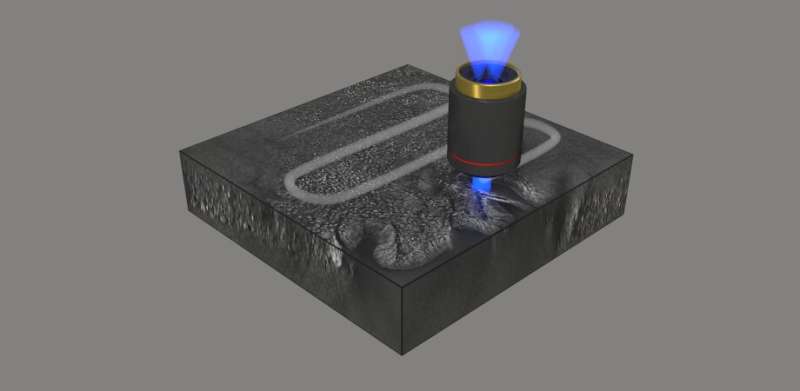
Outmoded methods in histopathology are on the overall restricted to thin specimens and require chemical processing of the tissue to supply sufficiently high distinction for imaging, which slows the approach. A recent are accessible histopathology eliminates the need for chemical staining and permits high-resolution imaging of thick tissue sections. As reported in Developed Photonics, a international research group currently demonstrated a 3D mark-free quantitative fragment imaging approach that uses optical diffraction tomography to form volumetric imaging records. Automatic stitching simplifies the image acquisition and prognosis.
Optical diffraction tomography
Optical diffraction tomography is a microscopy approach for reconstructing the refractive index of a tissue pattern from its scattered self-discipline photographs bought with diversified illumination angles. It permits mark-free high distinction visualization of clear samples. The complex scattered self-discipline transmitted thru the pattern is first retrieved the employ of off-axis holography, then the scattered fields bought with diversified attitude of illuminations are mapped in the Fourier dwelling enabling the reconstruction of the pattern refractive index.

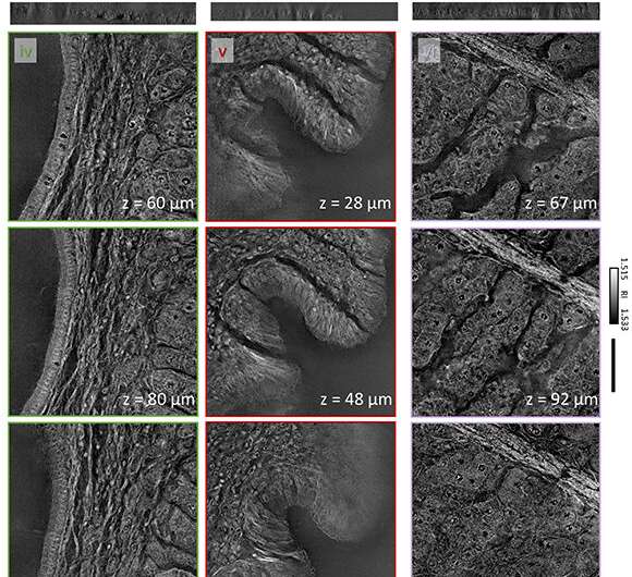
A acknowledged limitation of optical diffraction tomography is thanks to the complex distribution of refractive indexes, which leads to foremost optical aberration in the imaging of thick tissue. To conquer this limitation, the group ancient digital refocusing and automatic stitching, enabling volumetric imaging of 100-um-thick tissues over a lateral self-discipline of inquire of of two mm x 1.75 mm while sustaining a high resolution of 170 nm x 170 nm x 1400 nm. They demonstrated that simultaneous visualization of subcellular and mesoscopic structures in varied tissues is enabled by high resolution mixed with a wide self-discipline of inquire of.
Snappily, excellent histopathology
The researchers demonstrated the strategy of their unique manner by imaging a diversity of assorted most cancers pathologies: pancreatic neuroendocrine tumor, intraepithelial neoplasia, and intraductal papillary neoplasm of bile duct. They imaged millimeter-scale, unstained, 100-μm-thick tissues at a subcellular 3D resolution, which enabled the visualization of particular person cells and multicellular tissue architectures, a connected to photographs bought with primitive chemically processed tissues. Per YongKuen Park, researcher on the Korea Developed Institute of Science and Skills and senior writer on the understand, “The pictures bought with the proposed manner enabled sure visualization of assorted morphological parts in the diversified tissues allowing for recognition and diagnosis of precursor lesions and pathologies.”
Park notes that further research is foremost, but the outcomes suggest gigantic most likely for fleet, excellent histopathology all over surgical operation: “Extra research is foremost on pattern preparation, reconstruction speed, and mitigation of a pair of scattering. We inquire of optical diffraction tomography to supply quicker and more true diagnostics in histopathology and intraoperative pathology consultations.”
Extra records:
Herve Hugonnet et al, Multiscale mark-free volumetric holographic histopathology of thick-tissue slides with subcellular resolution, Developed Photonics (2021). DOI: 10.1117/1.AP.3.2.026004
Quotation:
Holographic histopathology permits fleet, true diagnostics (2021, April 30)
retrieved 2 Would possibly possibly possibly perhaps 2021
from https://phys.org/records/2021-04-holographic-histopathology-permits-fleet-true.html
This document is arena to copyright. Aside from any lovely dealing for the motive of non-public understand or research, no
fragment shall be reproduced with out the written permission. The lisp is equipped for records functions perfect.